Home »
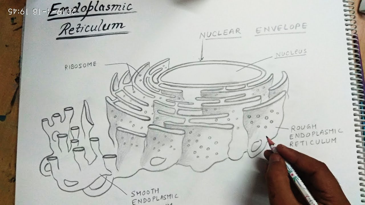
» Animal Cell Smooth Endoplasmic Reticulum Drawing / How To Draw Endoplasmic Reticulum Step By Step For Beginners Youtube / Endoplasmic reticulum (er) is the tubular membrane inside the cytoplasm of the cell.
Animal Cell Smooth Endoplasmic Reticulum Drawing / How To Draw Endoplasmic Reticulum Step By Step For Beginners Youtube / Endoplasmic reticulum (er) is the tubular membrane inside the cytoplasm of the cell.. It synthesizes lipids, phospholipids as in plasma membranes, and steroids. The cells of animals are the basic structural units for the wide. A labeled diagram of the animal cell and its organelles. Let's draw an animal cell:cell membranenucleus,mitochondriaendoplasmic reticulum,ribosomeschromatidsvacuoles andlysosomes!oh and let's not forget cytoplasm!l. The golgi body (yellow) modifies and packages proteins.
There are 13 main parts of an animal cell: It synthesizes lipids, phospholipids as in plasma membranes, and steroids. The er is a multifunctional organelle; A cell is the smallest unit of life; They help in the synthesis of proteins.
How To Draw Endoplasmic Reticulum Youtube from i.ytimg.com Illustration of organelles in an animal cell. Fusion of two haploid gametes to form a diploid zygote. Endoplasmic reticulum is an organelle found in both eukaryotic animal and plant cells. An animal cell diagram is a great way to learn and understand the many functions of an animal cell. The endoplasmic membrane consists of a network of membranous sacs called cisternae that branches off from the nuclear membrane. Both types consist of membrane enclosed, interconnected flattened tubes. Besides, the er has both smooth and rough surface. The er is a multifunctional organelle;
Also, ribosomes cover their surface.
Endoplasmic reticulum is an organelle found in both eukaryotic animal and plant cells. The synthesis of membrane lipids, membrane and secretory proteins, and the regulation of intracellular calcium are prominent among its array of functions. Begin labeling your organelles and other cell structures. Shop for smooth endoplasmic reticulum wall art from the world's greatest living artists. Fusion of two haploid gametes to form a diploid zygote. Lysosomes are the large ovals. Smooth endoplasmic reticulum( light blue) briefly describe the function of the cell parts. Imagenes fotos de stock y vectores sobre rough endoplasmic. The smooth endoplasmic reticulum, on the other hand, does not have ribosomes. The general appearance of the smooth endoplasmic reticulum is a membrane folded over on itself, a feature that greatly increases the surface area where metabolic processes can occur without taking up excess space within the cell. Cell nucleus and endoplasmic reticulum. At centre is the nucleus (transparent), which contains chromosomes (red) that hold the cell's genetic information. The er performs multiple functions in both plant and animal cells.
An animal cell diagram is a great way to learn and understand the many functions of an animal cell. See endoplasmic reticulum stock video clips. A plant cell diagram showing the smooth endoplasmic reticulum. Animal cell anatomy diagram structure with all parts nucleus. Imagenes fotos de stock y vectores sobre rough endoplasmic.
X0lrttwyxsvuxm from i.ytimg.com To draw a well labelled diagram of plant and animal cells, the golgi apparatus is placed near the endoplasmic reticulum. The cells of animals are the basic structural units for the wide. Ribosomes are found freely in the cytoplasm of the cell or attached to the membranes of endoplasmic reticulum. Cell nucleus and endoplasmic reticulum. The smooth endoplasmic reticulum has a tubular form. It's functions include producing and digesting lipids and also membrane proteins. An animal cell diagram is a great way to learn and understand the many functions of an animal cell. Eukaryotic cells are larger, more complex, and have evolved more recently than prokaryotes.
Cell nucleus and endoplasmic reticulum.
Rough er contains attached ribosomes while smooth er does not. Choose your favorite smooth endoplasmic reticulum photographs from 105 available designs. A cell's endoplasmic reticulum (er) contains a network of tubules and flattened sacs. Cell membrane, nucleus, nucleolus, nuclear membrane, cytoplasm, endoplasmic reticulum, golgi apparatus, ribosomes, mitochondria, centrioles, cytoskeleton, vacuoles, and vesicles. There are 13 main parts of an animal cell: 20% off all wall art! Both types consist of membrane enclosed, interconnected flattened tubes. The smooth endoplasmic reticulum, or smooth er, is an organelle found in both animal cells and plant cells. Where, prokaryotes are just bacteria and archaea, eukaryotes are literally everything else. Fusion of two haploid gametes to form a diploid zygote. The smooth endoplasmic reticulum, on the other hand, does not have ribosomes. The er is a multifunctional organelle; Transportation of food material, water and minerals in the body of a living organism.
Color a typical animal cell according to the directions to learn the main structures and organelles found in the cell. Animal cell anatomy diagram structure with all parts nucleus smooth rough endoplasmic reticulum cytoplasm golgi apparatus. Where, prokaryotes are just bacteria and archaea, eukaryotes are literally everything else. The golgi body (yellow) modifies and packages proteins. At centre is the nucleus (transparent), which contains chromosomes (red) that hold the cell's genetic information.
Structure Of The Rough And Smooth Endoplasmic Reticulum Illustrations from s.yimg.com Choose your favorite smooth endoplasmic reticulum photographs from 105 available designs. Choose your favorite smooth endoplasmic reticulum designs and purchase them as wall art, home decor, phone cases, tote bags, and more! Nucleus, mitochondria, membrane, centrosome, ribosome, smooth and rough endoplasmic difference between bacteria, animal and plant cells. The diagram, like the one above, will include labels of the major parts of an animal cell including the cell membrane, nucleus, ribosomes, mitochondria, vesicles, and cytosol. Illustration of organelles in an animal cell. They help in the synthesis of proteins. Plants and bacteria both have cell walls but they are shaped differently. Begin labeling your organelles and other cell structures.
From amoebae to earthworms to mushrooms, grass.
Its main functions are the synthesis of lipids, steroid hormones, the detoxification of harmful metabolic byproducts and the storage and metabolism of calcium ions within the cell. See endoplasmic reticulum stock video clips. They help in the synthesis of proteins. Eukaryotic cells are larger, more complex, and have evolved more recently than prokaryotes. The synthesis of membrane lipids, membrane and secretory proteins, and the regulation of intracellular calcium are prominent among its array of functions. An animal cell diagram is a great way to learn and understand the many functions of an animal cell. Imagenes fotos de stock y vectores sobre rough endoplasmic. Animal cell anatomy diagram structure with all parts nucleus smooth rough endoplasmic reticulum cytoplasm golgi apparatus. It is another membranous organelle near the endoplasmic reticulum as its primary function packs the proteins coming from the endoplasmic reticulum into vesicles before sending them to the required site or location in the cell. The golgi body (yellow) modifies and packages proteins. It is a subset of the endomembrane system of the endoplasmic reticulum. Endoplasmic reticulum images stock photos vectors. Nucleus, mitochondria, membrane, centrosome, ribosome, smooth and rough endoplasmic difference between bacteria, animal and plant cells.


Tidak ada komentar:
Posting Komentar